



Macular Hole Vitrectomy
Macular Hole Vitrectomy Surgery in Texas
Advanced Macular Hole Vitrectomy in Texas Restores Central Vision
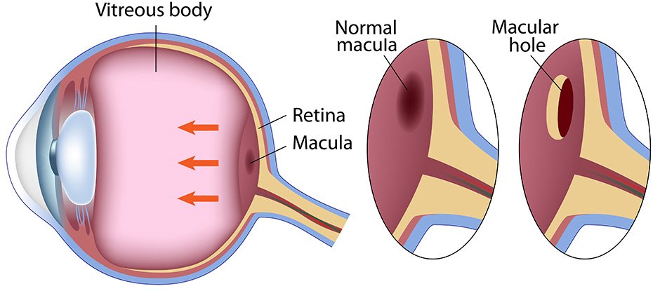
A macular hole is a full-thickness defect in the middle region of the retina (macula), which is responsible for sharp, detailed vision. If this thin tissue develops a hole, patients may have blurred center vision, distorted images, or a black patch that interferes with reading, driving, and recognizing faces. The usual surgical procedure to repair a full-thickness retinal hole and improve visual function is vitrectomy with implantation of a gas bubble.
Texoma Retina offers macular hole vitrectomy surgery in Texas using modern microsurgical procedures. Our skilled retina experts offer tailored care to patients who are seeking macular hole vitrectomy in Texas, helping them achieve the best possible visual outcomes with quick diagnosis and treatment.

What Is a Macular Hole?
A macular hole is a small tear in the macula, the portion of the retina responsible for sharp, central vision. It is most often caused by age-related changes in the vitreous gel that tugs on the retina. As traction grows, a full-thickness defect of the macula may be created.
The patient will often observe:
- Dark or blurry region in the center of the eyesight
- Straight lines are bent or torn in two.
- Difficulty reading, identifying faces, or completing intricate work
- Side vision normal, center vision impaired
If you have these symptoms, it is crucial to be evaluated promptly because early diagnosis can boost the chances of successful therapy.
When Is Vitrectomy Recommended?
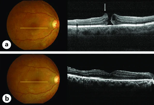
Vitrectomy for macular hole is suggested when an optical coherence tomography (OCT) scan shows a full-thickness macular hole, which usually happens in stages 2 through 4. Your retina expert can use OCT imaging to establish the size and severity of the hole and whether surgery is appropriate.
Patients needing advanced treatment for a macular hole benefit from early intervention since the sooner the surgery is performed after diagnosis, the better the visual recovery is usually, compared to late treatment.
How is the macular hole vitrectomy executed?
Macular hole vitrectomy surgery in Texas is usually done as an outpatient treatment with local anesthesia.
The retina surgeon does a standard three-port pars plana vitrectomy through tiny holes in the eye. To get rid of stickiness, the vitreous gel is carefully taken away, paying special attention to the area around the macula. A posterior vitreous detachment (PVD) can be safely caused if needed.
The internal limiting membrane (ILM) is then stained with a special dye like Brilliant Blue G or indocyanine green. This makes it easier to see. The surgeon carefully peels off this thin membrane around the macular hole with tiny tools to reduce friction and speed up healing.
A fluid-air exchange changes the eye’s natural fluid with air. Next, a long-lasting gas bubble like SF₆ or C₀F₈ is put in place. While the retina heals, the gas bubble softly presses the edges of the macular hole together. Lastly, the surgical sites are checked out. The small cuts usually heal on their own without stitches.
The goal of this advanced retina surgery for macular hole is to close the hole and recover central vision as much as possible.
What's the Point of Lying Face-Down?
Patients who have had surgery to repair a macular hole are often told to stay in a face-down position for a few days.
By keeping the gas bubble in this position, it stays in direct touch with the macula and helps seal the hole while the retinal tissue heals properly. Your surgeon will give you specific instructions about how long it will take based on the macular hole’s size and location.
Recovery After Surgery
It takes time for people to get better after macular hole treatment in Sherman, Texas.
Right away after surgery:
- As long as the gas bubble is there, vision will be fuzzy.
- The gas bubble temporarily makes it hard to see in the eye that is being treated.
- As the bubble naturally breaks down over a few weeks, things get better over time.
- During recovery, you may need to limit your activities.
- People should not fly until the gas bubble disappears for good.
Depending on how long the macular hole was there before surgery, most people notice their central vision getting better over a few weeks to a few months.
Success Rate and Expected Outcomes
Today’s macular hole surgery in Texas has excellent results, with 90–95% success rates for small and medium-sized holes.
Many retinal holes close up properly after surgery. The amount of vision improvement depends on the size of the hole, how long it was there before treatment, and the overall health of the retina. In general, getting treatment earlier leads to better long-term results and better visual recovery.
Safety and Risks
Vitrectomy for macular hole has some risks, like any surgery, but serious problems don’t happen very often.
Some possible risks are:
- Detachment of the retina
- Formation of cataracts
- A short-term rise in eye pressure
- Infection
- Incomplete hole closure
Your retina specialist can check on your healing, see how well the gas bubbles are absorbing, and make sure your retina stays healthy while you’re healing by seeing you for regular follow-up appointments.
Expert Macular Hole Care at Texoma Retina
Advanced macular hole vitrectomy in Texas is something that Texoma Retina does for people in Sherman, Allen, McKinney, Gainesville, and other North Texas cities. The fellowship-trained retina doctors at our center are experts at restoring central vision using cutting-edge vitrectomy techniques and the newest surgical tools.
We are happy to offer macular hole treatment in Sherman, Texas, and also help people from Durant, Oklahoma, and nearby towns. Our team of experienced retina specialists is dedicated to providing compassionate, high-quality care, whether you are looking for a retina specialist near Ardmore, Oklahoma, someone who is trusted by people all over Southern Oklahoma, or someone who is making the short drive from Durant.
If you notice a central blind spot, blurred vision, or picture distortion, you should see a doctor right away. When used correctly, advanced macular hole treatment can make it much easier to see. Set up a consultation right away to find out what the best way is to treat your eyes.
Frequently Asked Questions
Usually, vitrectomy surgery is used to fix a full-thickness macular hole. The internal limiting membrane is peeled off and the vitreous gel is taken out during the procedure. A gas bubble is then put inside the eye to help the hole close up as it heals.
The vitreous gel is taken out during a microsurgical process called vitrectomy to free up the macula from pressure. To help close the macular hole, the operator may peel back thin membranes and put a gas bubble inside the eye.
As the gas bubble slowly presses the macular hole's edges together, it helps the retinal tissue heal properly. Over time, the bubble gradually disappears, and the eye's own fluid takes its place.
Putting the face down helps the gas bubble stay in close contact with the macula. This makes it more likely that the hole will close properly and helps the body heal properly in the days following surgery.
Modern vitrectomy surgery can successfully close about 90–95% of small and medium-sized macular holes. Getting diagnosed and treated early usually gives people the best chance of seeing better.
Request an
Appointment
LET’S GET STARTED
Stay up to date on solutions and news
Opening Hours
Calls are taken 24/7 for emergencies
Available on Saturday and Sunday for Emergency only. Call for appointment
LOCATIONS AND DIRECTIONS