


Diagnostic Techniques
Diagnostic Techniques in Texas
Flourescin Angiography (FA)
Flourescin angiography is a diagnostic test used to help physicians identify eye conditions impacting circulation within the eye. The diagnostic testing lasts around 30 minutes.
Flourescin angiography begins first by dilating the eyes. An injection is given in the arm, which contains a yellow dye known as flourescin. The dye is injected within the arm, and the dye travels up to the eye. If there is any pathology within the eye, the flourescin dye will allow an Opthalmologist to clearly view the location of a damaged vein, artery, or other impacted structures within the eye. An ophthalmologist will use a specialized camera to clearly view the portions of the eye that may signify pathology.
No there are typically not any long-term effects of flourescin angiography. Typically symptoms subside within a minutes to about 24 hours after the dye is injected. When the dye is injected, some of it may come into contact with the skin. A slight burning sensation may occur, which will last for a few minutes, and quickly subside. When viewing objects, they could appear slightly darkened. This will not last more than a few minutes after the injection. Urine may appear a dark yellow or orange and will return to normal after 24 hours. This is typically due to the fact that the kidneys will filter the dye out of the body. Lastly, skin may appear somewhat yellow. Skin will return to yellow after a few hours. Very rarely, there may be a risk of an allergic reaction, which would immediately be treated.
Immediately after the procedure, sunglasses would be needed. Vision may appear blurry and exhibit sensitivity to light. A person may need a ride home, so it is advised to bring someone along to assist with transportation.
Optic Coherence Tomography (OCT)
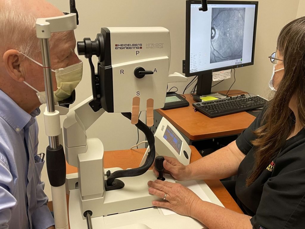
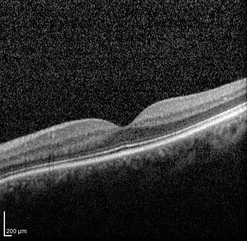
Optic Coherence tomography is a diagnostic technique used to take detailed images of the eye. Typically testing is completed within 10 minutes. An ophthalmologist may dilate the eyes, although some may choose not to. A patient typically will use a supporting cushion to allow the head to remain still. The machine will then scan the eyes, generating images. This is a noninvasive procedure.
This procedure is helpful in diagnosing several conditions that affect the retina, the macula, the vitreous, and lastly, the optic nerve.
There are typically no long-term risks regarding optic coherence tomography. Vision may be slightly blurred, but overall, no long-term risks have been known to occur.
Indocyanine Green Angiography
The Indocyanine Green Angiography is a diagnostic technique that is similar to Flourescin angiography. A Indocyanine Green dye is injected in the arm, and the dye eventually reaches the eye. This procedure is sometimes used along with Flourescin angiography because Indocyanine Green Angiography usually highlights the health of the choroid, while the Flourescin Angiography highlights pathology in the retina. A red infrared light camera is used to illuminate the dye and take images of the choroid and surrounding structures.
The Indocyanine Green Angiography does not typically have any long-term risks associated with it. The short-term side effects are similar to flourescin angiography.
Heidelberg Retinal Tomography
The Heidelberg retinal tomography is used to diagnose glaucoma. A laser is used to scan the retina, optic nerve, and determine the presence of signs of glaucoma. Oftentimes, early signs of cupping can begin, which is when there a loss of cells within the optic nerve, leading to symptoms of glaucoma.
Dilated Retinal Exam
This is an exam in which the pupil is dilated to be able to visualize the structures of the eye as well as the retina. The retina is assessed to determine any visual pathology
Visual Grid test/Amsler grid test
The Amsler Grid test is used to detect macular degeneration. Based on the grid, symptoms of macular degeneration may be present. While viewing the dot in the center, patients with macular degeneration may observe wavy lines, inability to view the corners of the grid, and seeings gaps in the grid.
A and B scans
The B scans are ultrasound scans are used to view the inner structures of the eye. The ultrasound waves help to visualize the individual structures of the eye and determine pathology. A scans help to view peripheral regions of lesions that may be present.
This procedure is helpful in diagnosing several conditions that affect the retina, the macula, the vitreous, and lastly, the optic nerve.
There are typically no long-term risks regarding optic coherence tomography. Vision may be slightly blurred, but overall, no long-term risks have been known to occur.
Comprehensive Retinal Diagnostics in Texas and Oklahoma
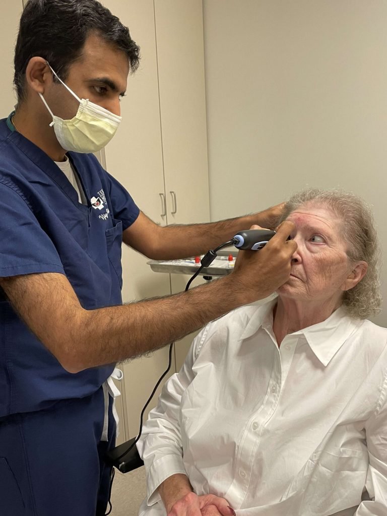
Retinal diagnostic testing provides ophthalmologists with the ability to identify, monitor, and manage illnesses of the retina, macula, vitreous, and optic nerve. Early retinal disease diagnosis is key to preventing loss of eyesight from illnesses such as diabetic retinopathy and macular degeneration.
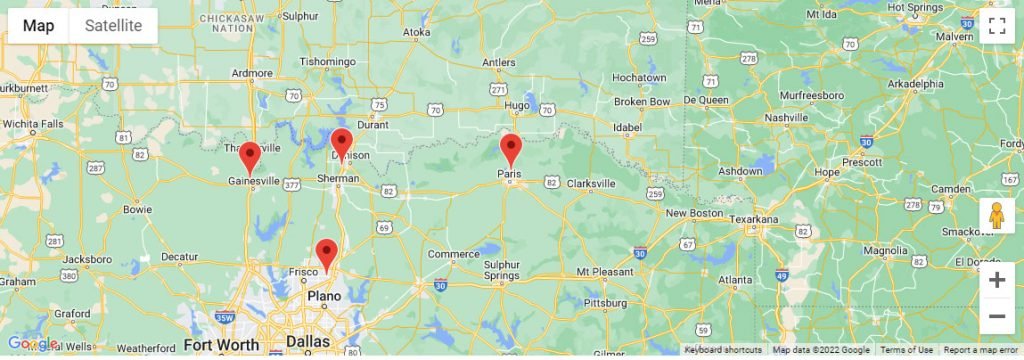
Advanced imaging can tell people who are looking for retina diagnostics in Texas a lot about the health of their retina and macula. A comprehensive examination may include numerous diagnostic tests, chosen based on the patient’s symptoms, medical history, and clinical results. Patients from Sherman, Denison, Gainesville, McKinney, Allen, and other North Texas cities may benefit from specialised retinal evaluation near home.
Patients from communities throughout Southern Oklahoma, including Durant, Calera, and surrounding areas, can also receive expert retinal evaluation when symptoms or existing eye diseases need additional research. Modern retina imaging center technology allows doctors to study the retinal structures in more depth and design a suitable diagnosis and treatment plan.
Retina Specialist Diagnostics
Retina specialist diagnostics use advanced imaging and specialty eye exams to look at conditions that are more complicated in the retina and vitreous. Tests may include OCT, angiography, retinal photography, ultrasound or other diagnostic procedures, depending on the clinical condition.
Retinal specialist diagnostics aren’t just about making an image; they’re also about giving clinically useful information that can help with choices about treatment and monitoring. The results may help doctors decide whether to treat with injections, laser therapy or surgery.
Advanced Eye Imaging in North Texas and Southern Oklahoma
Patients seeking advanced eye imaging in Texas can benefit from diagnostic services designed to help with conditions affecting the retina and macula. Specialised images can help doctors find small changes in the body’s structure or blood vessels and set a standard for future comparisons.
Advanced eye diagnostic tests are available at Texoma Retina in Sherman, Allen, McKinney, Gainesville, and other North Texas cities nearby. Our fellowship-trained retina specialists use cutting-edge imaging technology to make sure they make the right diagnosis and plan the right treatment.
Advanced diagnostic imaging can make it easy for people from Oklahoma who are travelling to get specialised retinal care to check out retinal problems in the Texoma region. Imaging tests can be used to check out patients from Durant and the southern Oklahoma towns nearby, based on their symptoms and clinical needs.
Having access to advanced eye imaging services in Texas can be very helpful when a thorough evaluation of the retina is needed for ongoing care or planning of treatment.
Why Retinal Diagnostics Matter?
Early diagnosis lets people get care before they lose their sight permanently. Many diseases that affect the retina don’t have any signs at first. Eye conditions caused by diabetes and getting older need to be checked on a regular basis.
Finding out more about the changes in a person’s eyes can also be helpful if they notice new floaters, flashes of light, distortion, blind spots, or sudden changes in their vision. Quick review may help find conditions that need medical help right away.
When to Schedule a Retinal Evaluation?
Early testing can help keep long-term damage from happening if you notice changes in your vision. Set up a full eye exam with our team of specialists today.
New flashes, a sudden rise in floaters, a shadow that looks like a curtain, distorted central vision, or a sudden loss of sight should all be checked out right away. A thorough check and the right imaging tests can help find out if there is a problem with the retina and whether treatment or ongoing monitoring is needed.
Request an
Appointment
Opening Hours
Calls are taken 24/7 for emergencies
Available on Saturday and Sunday for Emergency only. Call for appointment
LOCATIONS AND DIRECTIONS





Request an
Appointment
Opening Hours
Calls are taken 24/7 for emergencies
Available on Saturday and Sunday for Emergency only. Call for appointment
LOCATIONS AND DIRECTIONS