



Laser Peripheral Iridotomy (LPI)
Laser Peripheral Iridotomy (LPI) Treatment in Texas
Author: Aanya Roy and Aashna Bhakkad
Purpose: A laser procedure that creates a small hole in the peripheral iris (the colored part of the eye) to improve the flow of aqueous fluid between the anterior and posterior chambers. It is primarily used to prevent or treat angle-closure glaucoma by relieving pressure buildup behind the iris.
Understanding Angle-Closure Glaucoma
Angle-closure glaucoma is caused by a blockage of the drainage angle between the iris and cornea, which stops fluid from draining normally from the eye. This blockage can lead to a sudden increase in pressure in the eye and can cause lasting damage to eyesight if left unchecked.
Some people typically have narrow drainage angles, which means they may be more likely to have an angle close. In some cases, an acute angle-closure attack can cause significant eye pain, blurry vision, halos around lights, headaches, nausea, and a rapid rise in eye pressure. If you have any of these symptoms get medical help right away.
When LPI Eye Procedure is needed:
- Narrow or occludable angles detected during eye exam
- History or signs of acute angle-closure glaucoma
- High risk for angle closure based on family history or eye anatomy
- As a preventative treatment in the fellow eye of a patient with one affected eye
- When intraocular pressure (IOP) spikes due to blocked fluid flow
How Does Laser Peripheral Iridotomy Work?
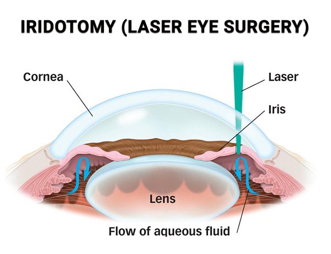
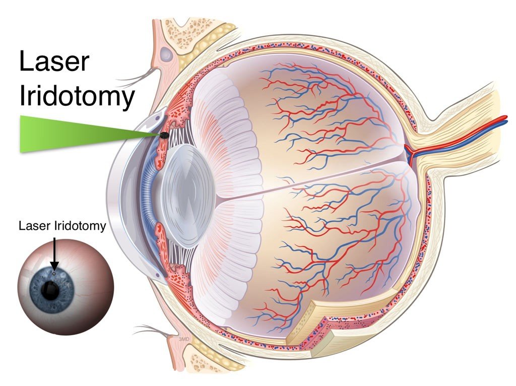
LPI makes a tiny hole in the peripheral iris that allows fluid to flow differently. This equalizes pressure between the front and back chambers of the eye and minimizes the chance of angle closure.
The laser iridotomy procedure permits the aqueous fluid to flow through the newly generated opening rather than getting trapped behind the iris. The technique may help to open or stabilize the drainage angle in suitable patients by enhancing fluid flow.
Procedure:
Setting: Outpatient procedure in an ophthalmologist’s office
Time: Usually 5–10 minutes per eye
Laser Used: YAG laser or argon laser, sometimes in combination
Function: Creates a new channel for fluid to flow through the iris, bypassing the pupil to reduce or prevent pressure buildup.
Steps:
- Numbing drops are applied to the eye
- Pupil may be constricted using a miotic agent (e.g., pilocarpine) to stretch the iris and reduce risk of bleeding
- Patient places head on the laser machine’s headrest
- A special contact lens with gel is placed on the eye to stabilize and focus the laser
- The laser is aimed at the peripheral iris (usually superior—under the upper eyelid for better cosmetic results)
- Laser pulses create a tiny full-thickness hole in the iris (0.2–0.5 mm)
- The contact lens is removed and the eye is rinsed
- Intraocular pressure is checked shortly after
- Anti-inflammatory eye drops (e.g., steroids or NSAIDs) are prescribed for a few days
What to Expect After LPI Eye Procedure?
- Temporary blurry vision, mild discomfort, redness, or light sensitivity
- Eye pressure may fluctuate—follow-up checks are important
- Most return to normal activities same day but should rest
- Avoid rubbing the eye and water exposure for 24–48 hours
Benefits
- Prevents or stops acute angle-closure attacks
- Reduces risk of vision loss from high eye pressure
- Quick, outpatient procedure with minimal recovery
- May avoid the need for emergency glaucoma surgery
- Provides long-term protection for angle-closure risk
Risks (Mostly Mild & Temporary)
- Inflammation (treatable with drops)
- Blurry vision, glare, or light sensitivity
- Temporary IOP spike
- Rare: bleeding, corneal edema, lens damage, or need for retreatment
Recovery Following LPI
Most patients resume normal activities within a day. You may experience temporary symptoms such as slight blurred vision, light sensitivity or eye inflammation. Follow-up visits are required to confirm that the opening is still patent and the drainage angle is still open.
Patients should follow the advice of their ophthalmologist concerning drugs and follow-up examinations. The LPI treatment gives another channel for fluid to drain. But glaucoma and other eye-pressure disorders may still need to be treated.
LPI as Part of Glaucoma Care
Laser Peripheral Iridotomy LPI is a well-known eye laser procedure for glaucoma that can be prescribed as a part of a complete angle closure glaucoma treatment plan. It may also be a significant narrow-angle glaucoma treatment for patients identified as being at risk.
Before suggesting laser peripheral iridotomy treatment in Texas to a patient, an ophthalmologist can look at the drainage angles, eye pressure, iris anatomy, and overall risk. LPI is not suitable for all glaucoma patients; hence, it is important to evaluate each patient individually.
At Texoma Retina & Glaucoma, patients are able to have a complete evaluation to see if LPI or any other glaucoma laser treatment is right for their situation. Early assessment can uncover narrow angles and outline the ideal way to prevent angle closure glaucoma and preserve vision in the long term.
Citations:
Gustianty, Elsa. “Evaluating the Effectiveness of Laser Peripheral Iridotomy on Angle-Closure Diseases: The Role of Spectral Domain Anterior Segment Optical Coherence Tomography (SD AS-OCT) in Indonesia Tertiary Eye Hospital.” The Open Ophthalmology Journal, vol. 19, no. 1, 26 Feb. 2025, openophthalmologyjournal.com/VOLUME/19/ELOCATOR/e18743641337429/FULLTEXT/.
IM Miguel, Ana, and André Borges Silva. “Laser Peripheral Iridotomy – EyeWiki.” Eyewiki.org, 22 Dec. 2023, eyewiki.org/Laser_Peripheral_Iridotomy? Accessed 1 Aug. 2025.
Joy, Rebecca. “Will Medicare Cover Glaucoma Care?” Healthline, Healthline Media, 14 Sept. 2020, www.healthline.com/health/medicare/does-medicare-cover-glaucoma?
Kelly, Sonia . “Laser Peripheral Iridotomy (LPI) | Narrow-Angle Glaucoma Treatment.” Allaboutvision.com, 14 Apr. 2025, www.allaboutvision.com/treatments-and-surgery/vision-surgery/glaucoma/laser-peripheral-iridotomy/?
Paretts, Susan. “Glaucoma Treatments, Costs and Insurance Explained.” Carecredit.com, 13 Dec. 2021, www.carecredit.com/well-u/health-wellness/glaucoma-surgery-cost-financing/.
Frequently Asked Questions
LPI is primarily conducted to lessen the risk of angle closure by providing another route for aqueous fluid to traverse the iris. It may be used for patients with narrow or occludable angles, angle-closure glaucoma, or greater risk of an acute episode.
Topical anesthetic drops are given to numb the eye beforehand. Most patients handle LPI well, but they may suffer temporary discomfort, pressure, light sensitivity, or irritation during or following treatment.
An LPI technique itself usually takes 5 to 10 minutes per eye. Preparation, pressure checks, and post-procedure instructions can make the whole session longer.
Laser peripheral iridotomy may be useful in chosen patients to decrease the probability of a pupillary-block-related angle closure. But it is contingent upon the underlying eye architecture and the type of glaucoma, and ongoing monitoring may be needed.
Risks include transient inflammation, impaired vision, sensitivity to light, glare, and a temporary increase in ocular pressure. Rare consequences may be hemorrhage, corneal edema, lens damage, or the need for retreatment.
Request an
Appointment

LET’S GET STARTED
Stay up to date on solutions and news
Opening Hours
Calls are taken 24/7 for emergencies
Available on Saturday and Sunday for Emergency only. Call for appointment
LOCATIONS AND DIRECTIONS