



Retinal Detachment Vitrectomy
Retinal Detachment Vitrectomy Treatment in Texas
Advanced Retinal Detachment Vitrectomy Treatment in Texas
Retinal detachment is a serious eye ailment and requires prompt medical intervention. This occurs when the retina pulls away from the supporting tissue beneath it, cutting off its blood supply and resulting in sudden, perhaps permanent vision loss if not addressed. Vitrectomy surgery is one of the most successful procedures in reattaching the retina and restoring anatomical stability by eliminating vitreous traction and mending retinal tears.
At Texoma Retina we perform retinal detachment vitrectomy Treatment in Texas using modern microsurgical techniques. Our skilled retina surgeons perform retinal detachment surgery in Texas for patients with simple and complex retinal detachments to preserve eyesight through early diagnosis and skillful treatment.
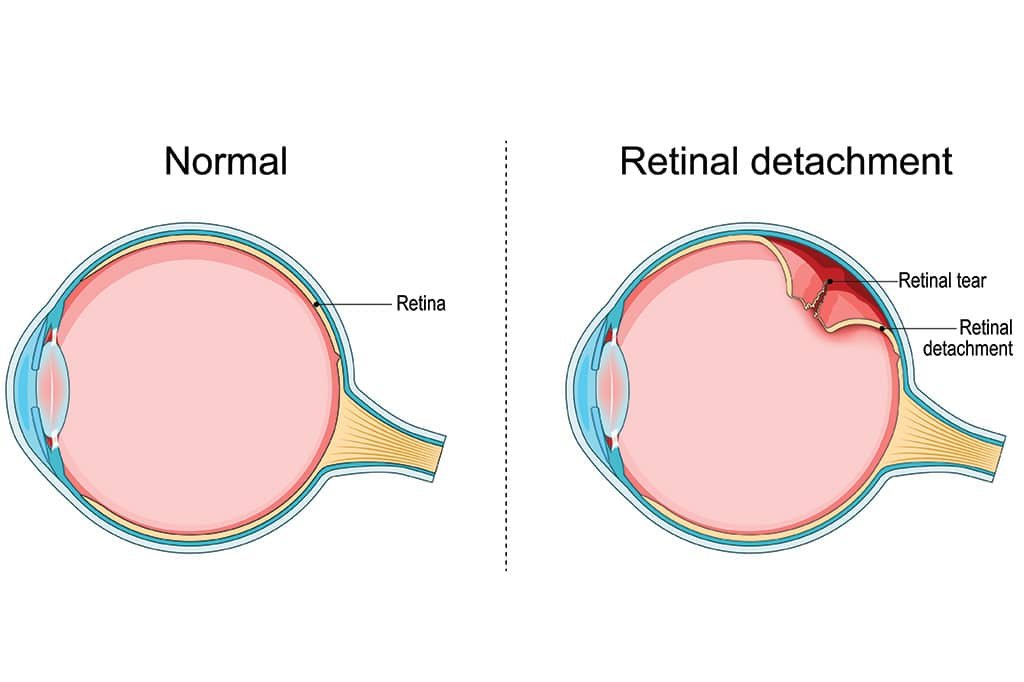
What Is Retinal Detachment?
The retina is the light-sensitive nerve tissue lining the back of the eye. When it comes away from its natural position, it cannot work properly, and vision is lost quickly.
There are different types of retinal detachment, for example:
Rhegmatogenous retinal detachment—when a tear in the retina permits fluid to pool underneath the retina.
Tractional retinal detachment—scar tissue pulls the retina away and is typically related to diabetic eye illness.
Macula-on vs. Macula-off detachment—the center retina (macula) is either still attached or has already detached and has a substantial impact on the visual outcomes.
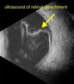
Retinal detachment is a true ophthalmologic emergency. If you see flashes of light, a rapid rise in floaters, or a curtain-like shade over your vision, you need to see someone right once. If not treated immediately, it can result in permanent blindness.
When Should You Get a Vitrectomy?
Vitrectomy for retinal detachment is recommended for:
- Large or complicated retinal detachments
- Macula-on (macula-threatening) detachments
- Retinal detachments with macula off
- Retinal detachments that are not responding to scleral buckle or pneumatic retinopexy
- Tractional retinal detachments from scar tissue
The sooner the surgery, the better the possibility of saving vision and preventing irreparable damage to the retina.
How does a retinal detachment vitrectomy perform?
Patients usually get local anesthesia and sedation for retinal detachment repair surgery, but general anesthesia may be needed for more complicated cases.
Fine 23-, 25-, or 27-gauge instruments are used to make three small cuts in the pars plana to start the process. An infusion cannula keeps the eye’s internal pressure steady while the vitreous gel is carefully taken out to stop pulling on the retina.
If needed, the surgeon causes a posterior vitreous detachment (PVD) and carefully shaves the vitreous base to stop it from pulling on the retinal tears in the future. A watching system with a wide field of view helps find all retinal breaks.
The subretinal fluid is then drained, which is often done with the help of perfluorocarbon liquid (PFCL). This makes the retina flatten against the back of the eye. Laser photocoagulation or cryotherapy is used to seal retinal tears and make a permanent connection.
Lastly, a tamponade is placed inside the eye:
- Gas bubble (SF₆ or C₃F₈): Gives temporary support from the inside while the body heals.
- Silicone oil: It is used when support is needed for a longer time or when the patient can’t keep up with their positioning needs.
Once it is confirmed that the retina is securely attached, the tools are taken out and the wounds that heal themselves are carefully looked over. This new retinal detachment treatment is meant to reattach the retina and stop further vision loss.
Recovery After Surgery
How quickly you recover depends on whether bubbles of gas or silicone oil were used during surgery.
Patients may be told to keep their heads in certain positions so that the gas bubble stays against the fixed retina. After surgery, vision is often blurry at first, especially if there is still a gas bubble in the eye.
As the body heals, the gas bubble slowly goes away, and over the course of weeks or months, vision slowly gets better. Your retina specialist will give you specific instructions about what activities you can and can’t do, when you will need follow-up visits, and when you can return to normal activities when it is safe.
Success Rate and Risks
The main success rate of modern retinal detachment vitrectomy in Texas is about 85–90%. However, depending on how complicated the detachment is, some patients may need more than one procedure.
Some possible risks are:
- Formation of cataracts
- Recurrent detachment of the eye
- Tears in the retina
- Getting sick
- Having high eye pressure
- Floaters during recovery
How well things go depends a lot on how quickly surgery is done after the retina separates. Anatomical and visual outcomes are much better when treatment starts early.
Expert Emergency Retina Care at Texoma Retina
Texoma Retina offers retinal detachment treatment in Sherman Texas, and emergency retinal care to patients in Sherman, Allen, McKinney, Gainesville, and other North Texas cities. Our fellowship-trained retina surgeons are experts in sophisticated retinal reattachment surgeries employing the latest surgical technology.
We are delighted to serve patients from Durant, Oklahoma, and the neighboring towns, and we are glad to care for patients seeking a retina specialist near Durant Oklahoma. Located only a short drive from Durant, people throughout Southern Oklahoma trust our team to provide fast, compassionate care when every minute matters.
If you notice flashes of light, a shower of floaters, or a veil over your vision, seek emergency retina surgery immediately. Early assessment and management could save your sight.
Frequently Asked Questions
Yes. A retinal detachment is a medical emergency. Prompt assessment and management are critical, as delay in treatment might lead to permanent visual loss or blindness.
In a retinal detachment vitrectomy, the vitreous gel is removed to reduce traction, retinal tears are sealed with laser or cryotherapy, and a gas bubble or silicone oil is injected in the eye to encourage healing of the retina.
Retinal detachment may be due to a retinal tear (rhegmatogenous detachment) , scar tissue pushing on the retina (tractional detachment) , eye trauma, previous eye surgery, or symptoms of severe diabetic eye disease.
If the portion of the retina that provides detailed vision detaches, this is called a macula-off retinal detachment. This disorder often results in considerable vision loss and requires prompt surgery to optimize visual recovery.
Recovery depends on the severity of detachment and the type of tamponade utilized. Postoperative head positioning may be needed for patients and vision normally recovers gradually over weeks to months, with regular follow-up treatment.
Request an
Appointment
LET’S GET STARTED
Stay up to date on solutions and news
Opening Hours
Calls are taken 24/7 for emergencies
Available on Saturday and Sunday for Emergency only. Call for appointment
LOCATIONS AND DIRECTIONS