



GATT surgery in Sherman Texas
Author: Aashna Bhakkad
GATT surgery in Sherman Texas is a new type of minimally invasive glaucoma surgery (MIGS) that reduces intraocular pressure (IOP) by enhancing the eye’s natural drainage system. In GATT, a suture or microcatheter is passed via Schlemm’s canal and utilized to open the trabecular meshwork surrounding the angle. This permits the aqueous humor to drain more efficiently through the eye’s natural drainage mechanism. GATT is generally utilized for chosen patients with open-angle glaucoma and may be done alone or in conjunction with cataract surgery.
Patients interested in GATT Surgery for Glaucoma in Sherman TX can have a thorough glaucoma evaluation to see if the operation is right for them. For this kind of minimally invasive glaucoma surgery in Sherman TX, you don’t need a normal drainage implant or an external filtering bleb.
What is Gonioscopy-Assisted Transluminal Trabeculotomy (GATT)?
One type of minimally invasive glaucoma surgery (MIGS) that lowers intraocular pressure (IOP) is Schlemm’s canal surgery. This works by opening the eye’s natural drainage system from the inside out. With the help of a gonioscopic view, the surgeon puts a suture or microcatheter through the whole canal. They then tear or remove the inner wall of the trabecular meshwork. This treatment works best for people with open-angle glaucoma and is meant to keep the eye’s natural structure while lowering risks and shortening recovery time.
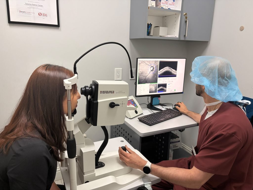
How does GATT work?
Additionally, GATT improves the flow of aqueous humor by making a circumferential opening in Schlemm’s canal. This lets fluid go around the diseased trabecular meshwork. A small goniotomy is made in the nose angle with a surgical lens, and the canal is threaded with a flexible suture (like 5-0 Prolene) or an illuminated microcatheter. The suture is pulled through, tearing the inner wall and opening the whole tube once the full 360° is created. This lowers IOP without using devices or sending fluid outside the body.
Who is a good Candidate?
- Primary open-angle glaucoma (mild to moderate stages)
- Juvenile or congenital glaucoma
- Pseudoexfoliative or pigmentary glaucoma
- Patients with prior failed MIGS but no major scarring
- Those undergoing combined cataract surgery
Not recommended for patients with: - Angle-closure glaucoma
- Heavily scarred angles or extensive synechiae
- Prior filtering surgeries that disrupted Schlemm’s canal
Steps During the Procedure
- Local anesthesia and mild sedation are administered
- A goniotomy is performed under gonioscopic view to access Schlemm’s canal
- A suture or microcatheter is threaded circumferentially (360°) through the canal
- Once full passage is achieved, the instrument is pulled through to unroof the trabecular meshwork
- The anterior chamber is flushed, and viscoelastic is removed
- Antibiotic and anti-inflammatory drops are given postoperatively
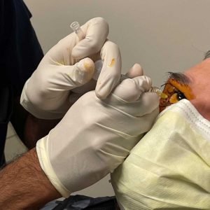
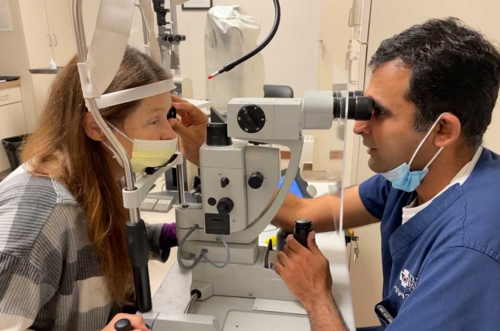
What to Expect Before, During, and After
- Before: Standard pre-op eye exam and imaging; discontinue contact lenses; fasting may be required
- During: The procedure takes about 30–45 minutes and is performed in an outpatient setting
- After: Mild irritation, redness, and blurred vision are common for a few days
Eye drops (steroids and antibiotics) will be used for 1–4 weeks.
Follow-up visits monitor IOP and healing. Return to normal activity in 1–2 weeks.
Is GATT Safe?
Yes. GATT has a favorable safety profile, especially compared to traditional surgeries like trabeculectomy:
- No external bleb or implant
- Preserves conjunctiva for future surgeries
- Lower risk of hypotony and infection
- Can be repeated or converted to other procedures if needed
Complications
Although rare, some potential risks include:
- Hyphema (blood in the anterior chamber)—common but usually self-resolving
- Transient IOP spikes
- Incomplete trabeculotomy if Schlemm’s canal cannot be fully cannulated
- Scarring or failure requiring additional IOP-lowering treatments
- Inflammation or corneal edema (rare)
Benefits of GATT surgery in Sherman Texas
- Minimally invasive with no implant or foreign body
- Can be done in combination with cataract surgery
- Effective for various open-angle glaucomas
- Quick recovery and return to normal activities
- Preserves tissue for future surgical options
Temporary redness, hazy vision, inflammation, and light sensitivity may occur after GATT treatment for glaucoma near Durant Oklahoma. Follow-up appointments are needed to check IOP and recovery. Patients undergoing GATT treatment in Allen Texas, should follow their surgeon’s directions regarding drugs and activities.
Insurance and Coverage
Typically covered by Medicare and most private insurers when medically indicated
CPT Code: 65850 (for trabeculotomy); a separate code may apply for catheter-based GATT.
GATT vs. Traditional Glaucoma Surgery
GATT is different from trabeculectomy and tube-shunt surgery in that it does not develop an external filtering bleb or include a traditional drainage implant. However, the technique is influenced by the severity of the glaucoma and the target IOP. Advanced eye care in Texas involves individualized evaluation, not choosing surgery on the basis of location or kind of operation alone.
While looking for the best eye doctor in Texas, patients should think about how knowledgeable the doctor is about glaucoma, how they can diagnose the condition, what treatment options are available, and how they will continue to care for patients after surgery.
Citations:
Fellman, Ronald L., and Davinder S. Grover. “MIGS and the Conjunctiva: Preserving the Future.” Glaucoma Today, July–Aug. 2014,
https://glaucomatoday.com/articles/2014-july-aug/migs-and-the-conjunctiva-preserving-the-future.
Grover, Davinder S., et al. “Clinical Outcomes of Gonioscopy-Assisted Transluminal Trabeculotomy in Eyes with Primary Open-Angle Glaucoma.” British Journal of Ophthalmology, vol. 101, no. 6, June 2017, pp. 743–749.
https://bjo.bmj.com/content/101/6/743.
American Academy of Ophthalmology. Primary Open-Angle Glaucoma Preferred Practice Pattern®. 2020.
https://www.aao.org/preferred-practice-pattern/primary-openangle-glaucoma-ppp.
Frequently Asked Questions
GATT dilates the trabecular meshwork and promotes passage of aqueous humor to Schlemm’s canal and the natural outflow system.
Yes. GATT is often regarded as minimally invasive glaucoma surgery, as it allows access to and enhances the natural drainage channel.
Selected patients with open-angle glaucoma might be candidates after assessment of IOP, angle anatomy, glaucoma severity, and prior treatment.
Yes. GATT can be combined with cataract surgery in chosen patients.
The procedure is usually an outpatient treatment, and the length of the surgery will depend on the patient and the conditions of the surgery.