



Glaucoma Tube Shunt Surgery in Texas
Author: Aashna Bhakkad
What is Glaucoma Tube Shunt Surgery?
A glaucoma tube shunt (also called a glaucoma drainage implant or device) is a surgical procedure used to lower intraocular pressure (IOP) in patients with moderate to advanced glaucoma, especially those who have not responded well to medications or prior surgeries like trabeculectomy.
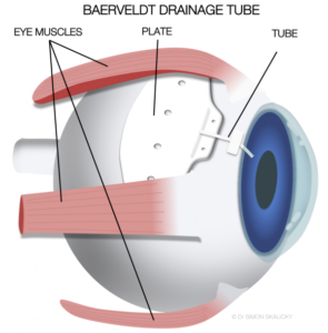
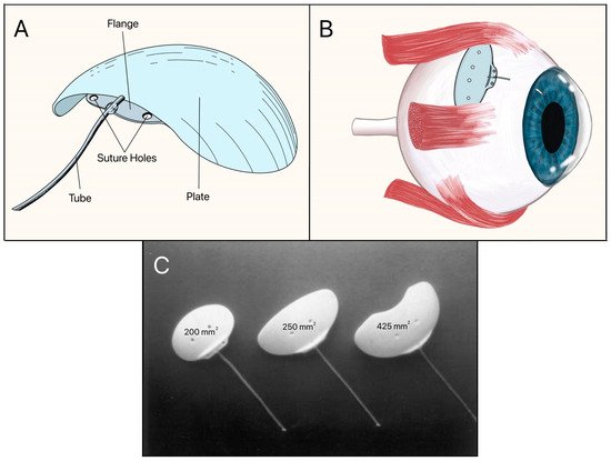
The procedure involves placing a tiny flexible tube (shunt) in the eye that drains excess aqueous humor to a small plate (reservoir) implanted under the conjunctiva, where the fluid is absorbed into surrounding tissue.
Why might you need tube shunt surgery?
If the pressure is not properly managed, glaucoma can damage the optic nerve over time. Some patients need incisional surgery, even though medicines, laser procedures, and minimally invasive glaucoma surgery may work for many. Glaucoma tube shunt surgery makes a different way for the aqueous fluid to leave the eye. It may be considered after other treatments have failed or when the case is very complicated.
Common Devices Used:
- Ahmed Glaucoma Valve (AGV)
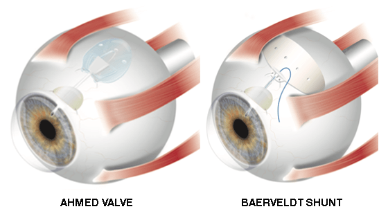
A valved implant that controls the flow of fluid out of the body and helps stop too much drainage right after surgery.
- Baerveldt Glaucoma Implant
A device without valves that has a bigger plate and is meant to lower pressure significantly.
- Molteno Implant
This well-known glaucoma drainage machine directs the aqueous humor toward a reservoir under the conjunctiva.
These vary in design (valved vs. non-valved), size, and drainage capacity but function similarly.
How it works:
- The surgeon implants a small plastic plate on the white part of the eye (sclera), usually under the upper eyelid.
- A thin tube is then inserted into the anterior chamber of the eye.
- Aqueous fluid flows through the tube to the plate, where it collects and is gradually absorbed, lowering IOP.
- In valved devices (like Ahmed), the valve helps regulate early postoperative pressure.
Who is a good candidate?
Tube shunts are often used in patients with:
- Failed trabeculectomy or other MIGS procedures
- Neovascular, uveitic, or traumatic glaucoma
- Congenital or pediatric glaucoma
- Extensive conjunctival scarring
- Uncontrolled IOP despite maximal medical therapy
They are generally reserved for moderate to severe cases or when angle-based surgeries are not viable.
What to Expect Before, During, and After:
- Before: Comprehensive glaucoma evaluation, possible medication adjustment, and lab work if general anesthesia is used
- During:
- Procedure typically takes 1–1.5 hours
- Performed under local or general anesthesia
- The tube and plate are secured to the eye and covered with donor tissue or scleral patch
- After:
- Eye drops (steroids, antibiotics) for 4–8 weeks
- Vision may be blurred for a few days to weeks
- Follow-up visits monitor pressure, tube position, and healing
- Final healing may take 6–12 weeks
Is Tube Shunt Surgery Safe?
Yes, though more invasive than MIGS, tube shunts are proven to be effective and durable, especially in complex or refractory glaucoma. Compared to trabeculectomy, they offer:
- Lower risk of early postoperative hypotony (especially with valved implants)
- Greater IOP control in high-risk eyes
- Long-term success rates with proper monitoring
Complications:
While generally safe, risks include:
- Hypotony (too low IOP)
- Tube blockage or migration
- Double vision (diplopia)
- Corneal endothelial cell loss or tube-cornea touch
- Blebitis or endophthalmitis (rare but serious infection)
- Chronic discomfort or irritation
Regular follow-up is essential to monitor tube position and eye pressure.
Benefits of Tube Shunts:
- Effective in eyes with prior failed surgeries
- Long-term pressure control
- Suitable for complex glaucomas (e.g., neovascular, uveitic)
- Can preserve remaining vision in advanced cases
- Valved models offer safer IOP regulation in early recovery
When standard treatment hasn’t lowered the eye pressure enough, a glaucoma drainage device can be very helpful.
Glaucoma Tube Shunt Surgery in North Texas and Southern Oklahoma
If you are a patient looking for glaucoma tube shunt surgery in Sherman Texas, you might want to get a full evaluation to establish whether a drainage implant or another treatment is appropriate for you. Depending on the diagnosis, treatment options may include drugs, laser therapy, minimally invasive glaucoma surgery, trabeculectomy, or tube shunt implantation.
If you are contemplating eye surgery for glaucoma in Texas, a glaucoma expert can analyze the damage of the optic nerve, IOP, past treatments, and overall eye health before recommending a procedure. Patients in Durant, Hugo, Atoka, Madill, Ardmore, and Coalgate can also consider glaucoma treatment surgery in Oklahoma while they explore specialized care in the surrounding North Texas area.
If you have tried medication, laser treatment, or less invasive treatments without enough control over the pressure, an incisional surgery specialist in North Texas can speak with you about whether a glaucoma drainage implant is right for you.
An incisional surgery specialist in North Texas can talk about if a glaucoma drainage implant is the right choice when drugs, laser treatment, or less-invasive treatments have not been effective in controlling pressure.
Insurance and Coverage:
- Covered by Medicare and most private insurers when medically necessary
- CPT Code: 66180 (Aqueous shunt to extraocular reservoir)
May require prior authorization depending on the insurer and device used.
Key Takeaway
Some surgeries, like glaucoma tube shunt surgery, can lower eye pressure when other methods don’t work. Depending on the patient, a glaucoma drainage device may offer an additional way for aqueous fluid to drain. It is important for people seeking glaucoma tube shunt surgery in Sherman, eye surgery for glaucoma in Texas, or specialized glaucoma treatment surgery in Oklahoma to get a full evaluation by a glaucoma specialist in order to obtain the best treatment.
Citations:
American Academy of Ophthalmology. Drainage Implant Surgery for Glaucoma. 19 Jan. 2022.
https://www.aao.org/eye-health/treatments/glaucoma-drainage-implant
National Eye Institute. Glaucoma Surgery. U.S. Department of Health and Human Services,
https://www.nei.nih.gov/learn-about-eye-health/eye-conditions-and-diseases/glaucoma/glaucoma-treatment.
Gedde, Steven J., et al. “Treatment Outcomes in the Tube Versus Trabeculectomy (TVT) Study after Five Years of Follow-up.” American Journal of Ophthalmology, vol. 153, no. 5, 2012, pp. 789–803.e2.
https://doi.org/10.1016/j.ajo.2011.10.024.
Ahmed Valve or Baerveldt Implant. Glaucoma Research Foundation.
https://glaucoma.org/ahmed-valve-or-baerveldt-implant/.
Levin, Leonard A., and Daniel M. Albert. Ocular Disease: Mechanisms and Management. Saunders Elsevier, 2010.
Frequently Asked Questions
It lowers IOP by sending aqueous humor to a storage under the conjunctiva through a small tube.
Candidates are chosen one by one. People with complex or uncontrolled glaucoma may be able to get it. The group includes some people who have already had surgery for glaucoma.
The Baerveldt doesn't usually have valves, but the Ahmed does. Their shapes and how the blood flows after surgery are different.
Like any other eye surgery, there are risks, including infection, bleeding, hypotony, corneal problems, and more. Before surgery, your ophthalmologist should talk to you about the risks and rewards.
According to the National Eye Institute, surgery for a glaucoma implant usually takes between one and two hours, but the length of time can change.